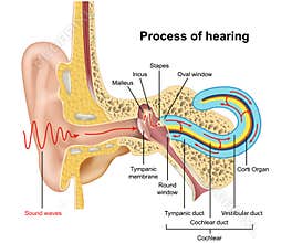
Ear - Anatomy. Frontal Section Through The Right External, Middle, And Internal Ear. Shown Are The Vestibular Labyrinth, Cochlear Labyrinth, Cochlea, Tensor Tympani Muscle, Auditory Tube,Tympanic Cavity, Tympanic Membrane, Temporal Bone, Auditory Ossicles, Auricle, External Acoustic Meatus, Mastoid Process,Styloid Process, Semicircular Canals, Incus, Malleus, Stapes, Vestibulocochlear Nerve
ID 169946896845 © Medicalartinc | Megapixl.com
CATEGORIES
Sharing is not just caring, it's also about giving credit - add this image to your page and give credit to the talented photographer who captured it.:
KEYWORDS
acoustic anatomy auditory aural auricle bone buds canals cavity cochlea cochlear deaf deafness external headphones hearing impediment incus labyrinth listen loud malleus mastoid meatus membrane muscle music nerve ossicles process quiet semicircular sound speech stapes styloid temporal tensor tube tympani tympanic vestibular vestibulocochlear


